


Dr Ng Yew Poh (Vincent)
Neurosurgeon


 Add as a preferred source on Google
Neurosurgeon
Being told that a scan has found a brain aneurysm can understandably bring many questions. Could it rupture? Does it need surgery? And if your specialist recommends monitoring it instead, is it really safe to wait?
Finding a brain aneurysm does not automatically mean that treatment is required. Some unruptured brain aneurysms may be monitored, while others may benefit from treatment.
What happens next depends on the individual aneurysm and the person living with it. A neurosurgeon will assess several factors to determine the risk it may pose and whether monitoring or treatment may be appropriate.
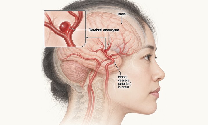
A brain aneurysm is a bulge that forms in a weakened area of the wall of a blood vessel in the brain. Risk factors include high blood pressure, cigarette smoking and family history. Brain aneurysms can occur in all age groups but have a higher incidence among those aged 40–60 and in women.
They are also more common than most people expect. Population studies estimate that roughly 3 in every 100 adults have an unruptured brain aneurysm, and the great majority will never know about it. Wider use of MRI and CT scanning means that more are now found incidentally, in people being scanned for headaches, dizziness, a head injury or as part of a health screening package.
An unruptured brain aneurysm has not burst and caused bleeding in the brain. Many unruptured aneurysms do not cause symptoms and may be discovered incidentally when a person undergoes a brain scan for another reason.
A ruptured brain aneurysm, on the other hand, causes bleeding in or around the brain and is a medical emergency.
Learn more about the symptoms and causes of brain aneurysms here.
Most unruptured aneurysms are first seen on one of three types of scan, and it is worth knowing which one you have, because they are not interchangeable.
A CT scan performed with contrast dye injected into a vein. It is quick and widely available and is usually the first test when someone presents acutely or when an aneurysm is suspected on a plain scan. It involves a dose of radiation.
An MRI-based study of the blood vessels. It involves no radiation and can often be performed without contrast, which makes it the usual choice for repeated monitoring over many years.
A catheter angiogram, in which a fine tube is passed from an artery in the wrist or groin and contrast is injected directly into the brain vessels. It gives the clearest picture of the aneurysm neck and the surrounding branches and remains the reference standard for planning treatment. Because it is an invasive procedure with a small risk of its own, it is reserved for situations where that extra detail will change the plan.
One practical point that surprises many patients: the measured size of the same aneurysm can differ by a millimetre or two between scan types, and even between projections on the same scan. This is part of why a single measurement is a weak basis for a decision, and why comparing like-for-like scans over time matters more than any one number.
No. Not every unruptured brain aneurysm needs to be treated immediately. In some people, the risk associated with treating an aneurysm may outweigh its estimated risk of rupture, and monitoring may therefore be recommended.
This is why discovering an aneurysm is usually followed by an individual assessment rather than an automatic recommendation for surgery.
The aim is to weigh two considerations:
This balance will be different for every patient.

There isn't a single measurement or characteristic that determines whether an aneurysm should be treated. Your specialist will look at the aneurysm itself as well as factors relating to your health and medical history.
These factors are not weighed by intuition alone. Validated tools such as the PHASES score bring six of them together — population, hypertension, age, aneurysm size, earlier subarachnoid haemorrhage and aneurysm site — to produce an estimated five-year risk of rupture. In the pooled international data behind PHASES, the average risk across all unruptured aneurysms studied was around 1.4% over one year and 3.4% over five years. A score of this kind is a starting point for a conversation rather than a verdict, but it turns a frightening unknown into a figure you can actually weigh against the risk of treatment.
The size of an aneurysm is one consideration when assessing its risk of rupture. However, size alone does not determine whether treatment is necessary.
A smaller aneurysm may still need closer assessment depending on its location, shape and other characteristics, while some aneurysms may be suitable for monitoring.
To give a sense of scale: in a large Japanese cohort of more than 6,000 unruptured aneurysms followed over time, the overall rupture rate was just under 1% per year. Aneurysms of 3 – 4mm ruptured at roughly 0.4% per year; those of 7 – 9mm carried around three times that risk, and those of 10mm or more considerably higher again.
Set against that, most aneurysms that do rupture measure less than 7mm. That is not a contradiction, small aneurysms are simply far more common, but it does mean that small is not the same as safe, which is exactly why size is never assessed on its own.
Where an aneurysm occurs within the brain's blood vessels can influence both its estimated risk of rupture and the options available for treating it.
Aneurysms arising from the anterior communicating and posterior communicating arteries, and those in the posterior circulation (the vertebral and basilar arteries and their branches), carry a higher risk of rupture than aneurysms of the same size elsewhere.
Your specialist will therefore consider the aneurysm's location together with its other characteristics rather than assessing any single feature in isolation.
The shape and surface contour of an aneurysm carry information that size alone does not.
A daughter sac (a small blister arising from the main dome), an irregular or lobulated outline, and a high dome-to-neck ratio are all associated with an increased risk of rupture. The neck also has a direct bearing on treatment: a narrow-necked aneurysm is usually straightforward to coil, whereas a wide-necked one may need a stent, a balloon or a different technique altogether.
If previous scans are available, your specialist may compare them to determine whether the aneurysm has changed in size or appearance over time.
Evidence of growth can affect how an aneurysm is managed.
An aneurysm that has demonstrably grown is regarded quite differently from one that has been stable for years, because documented growth is among the stronger signals that the wall is under stress. Changes of a millimetre have to be interpreted carefully, though, since some of that difference can come from the scan rather than the aneurysm — which is why serial imaging is ideally performed on the same modality each time.
Treatment itself carries risks, so your specialist will also consider your age, overall health and other medical conditions.
This helps determine whether the potential benefit of treating the aneurysm outweighs the risks associated with the procedure.
Certain health and lifestyle factors may also be considered, including high blood pressure and smoking.
A family history of brain aneurysms or aneurysmal rupture can also be relevant, particularly if close relatives have been affected.
Screening of unaffected relatives is generally offered where two or more first-degree relatives (a parent, sibling or child) have had a brain aneurysm or a subarachnoid haemorrhage, and to people with autosomal dominant polycystic kidney disease, in whom aneurysms are more common. A single affected relative does not usually warrant screening, but it is worth raising with your specialist.
“Almost every patient asks me first how big it is. Size does matter, but it is one of several things I weigh, including where the aneurysm sits, its shape, whether it has changed on previous scans, and the health and priorities of the person in front of me. My role is to bring those together, explain honestly what they mean, and reach a decision the patient feels settled about.”
—Dr Vincent Ng
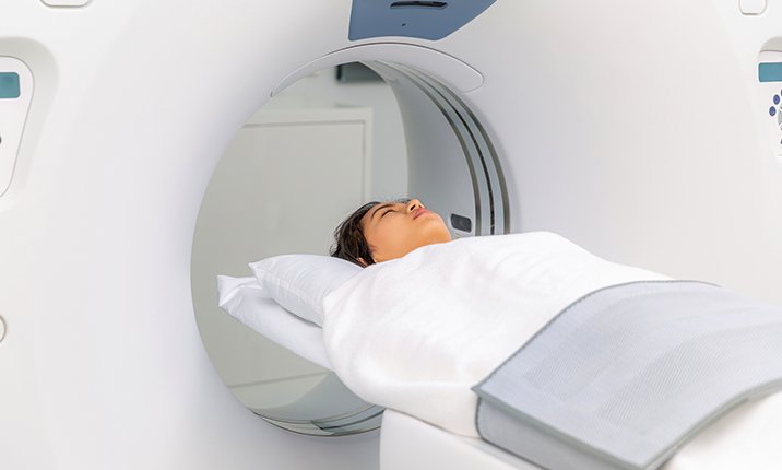
Being told that an aneurysm can be monitored rather than treated may initially sound worrying. However, monitoring does not mean ignoring the aneurysm.
Your specialist may recommend follow-up imaging to check whether its size or characteristics change over time.
The type and frequency of scans will depend on your individual condition, so there is no single monitoring schedule that applies to everyone.
In practice, a common pattern is a repeat scan at around six to twelve months to confirm the aneurysm is stable, then at progressively longer intervals if it remains unchanged. Some patients are eventually discharged from surveillance altogether; others, particularly younger patients, continue at intervals over many years.
Your specialist may also recommend addressing factors that can increase the risk of aneurysm growth or rupture, such as managing high blood pressure and stopping smoking.
If subsequent scans show changes to the aneurysm, your specialist can reassess whether continued monitoring or treatment is appropriate.
“When I tell a patient we will monitor an aneurysm, I want them to hear a plan rather than a postponement. Monitoring means agreed intervals for imaging, attention to blood pressure, support to stop smoking, and a clear understanding of what we are watching for and what would prompt us to act. Patients cope far better when they know exactly what is being done, and why.”
—Dr Vincent Ng
Treatment may be considered when your specialist determines that the potential risk posed by the aneurysm outweighs the risks associated with intervention.
The decision is individualised and may consider the aneurysm's:
As well as your:
If treatment is recommended, the next question is which approach is most suitable for your aneurysm.
There are several ways of treating a brain aneurysm. The aim is generally to prevent blood from entering the aneurysm and reduce the risk of it rupturing.
The appropriate approach depends on factors including the aneurysm's location, size and shape, as well as your overall health.
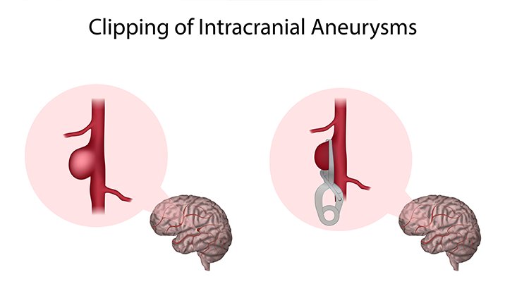
During surgical clipping, a neurosurgeon accesses the aneurysm and places a small clip across its neck. This prevents blood from flowing into the aneurysm.
Clipping is performed through a craniotomy under general anaesthesia. Once a clip is correctly placed across the neck, the aneurysm is sealed permanently and rarely needs further treatment.
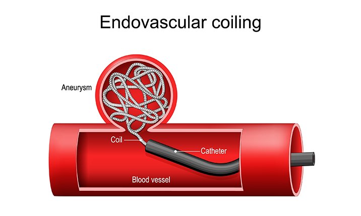
A type of endovascular embolisation, coiling treats the aneurysm from within the blood vessel.
A thin catheter is guided through an artery towards the aneurysm. Small coils are then placed within the aneurysm to reduce blood flow into it and promote clotting, effectively sealing it off from the circulation.
Because the aneurysm is reached through the blood vessels, the procedure does not require opening the skull.
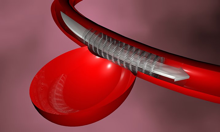
For certain aneurysms, a flow-diverting stent may be considered. (A type of endovascular embolisation)
Rather than filling the aneurysm itself, the device is placed within the affected blood vessel to redirect blood flow away from the aneurysm, allowing it to gradually close off.
Aneurysms with a wide neck can be difficult to coil on their own. In these cases, a balloon may be used temporarily to hold the coils in place, a stent may be placed across the neck of the aneurysm, or an intrasaccular device such as a braided mesh implant may be positioned inside the aneurysm sac itself.
Not every technique is suitable for every aneurysm. Which options are open to you is determined largely by the anatomy of the aneurysm and the vessel it arises from.
There is no universally "better" treatment for every brain aneurysm.
A specialist may consider factors such as the aneurysm's size, shape and location, the anatomy of the surrounding blood vessels, whether the aneurysm has ruptured, and the patient's overall health when recommending an approach.
Each treatment also has its own potential benefits and risks, which should be discussed with your specialist.
This is the part patients most often forget to ask about, and it matters for planning work and family life.
Most patients go home within a few days — typically a night or two after endovascular treatment (flow diversion or coiling), and a few days after clipping. Returning to everyday activity generally takes somewhere between one and a few weeks, depending on the procedure, the aneurysm treated, and your own rate of recovery. Your surgeon will give you a realistic timeframe for your situation before any decision is made.
Follow-up imaging is the usual practice after treatment.
It is however done more frequently and for a longer duration after coiling than after clipping. Your specialist will advise on driving, air travel, exercise, heavy lifting and, where relevant, pregnancy — most patients return to all of these activities, but the timing depends on the treatment and on your recovery.

Any treatment that leaves a stent or flow diverter inside the blood vessel normally requires a period of dual antiplatelet medication, typically aspirin together with a second agent started before the procedure and continued for months afterwards, often with aspirin continued long term. This carries a small bleeding risk of its own and needs to be planned around any other surgery or dental work. Clipping and standalone coiling do not require it. For some patients this is a decisive consideration, and it is worth raising early rather than after the decision is made.
It can be difficult to remember what to ask after receiving an unexpected diagnosis. Consider bringing these questions to your consultation:
If you are seeking another opinion, bringing your previous scan images and reports may also help your specialist understand how the aneurysm was discovered and assess its characteristics.

An unruptured brain aneurysm is different from a ruptured aneurysm.
When an aneurysm ruptures, bleeding can occur around the brain and urgent medical treatment is required.
A sudden, extremely severe headache, sometimes described as a thunderclap headache, is a characteristic warning sign of rupture. Other symptoms can include nausea and vomiting, neck stiffness, loss of consciousness, seizures, confusion or neurological changes.
In some patients, a smaller warning leak causes a sudden and unusually severe headache days or even weeks before a major rupture. This pain might settle after a while, and might be dismissed as a migraine. However, ignoring it at this juncture may potentially lead to patients missing the window to treat the aneurysm before the full rupture happens (and more brain damage has occurred). As such, any headache that reaches full intensity within seconds and is the worst you have ever experienced should be assessed urgently.
If you or someone around you develops symptoms that could indicate a ruptured brain aneurysm, seek medical attention immediately.
A brain aneurysm diagnosis can sound frightening, but it does not tell you by itself what will happen next.
Some aneurysms may require treatment, while others can be monitored. Understanding the difference requires an assessment of the aneurysm's characteristics alongside your individual health and risk factors.
“A diagnosis like this is frightening, and that fear is entirely understandable. What helps most is clarity — knowing what the aneurysm is, what risk it genuinely carries, and what can be done about it. In my experience, most patients find the way forward far more manageable once they have that.”
—Dr Vincent Ng
At Mount Elizabeth Hospitals, our neurosurgeons can assess your condition, discuss the management options available and help you understand the next steps in your care.
Yes. Some people with unruptured brain aneurysms may be monitored rather than treated immediately. Whether this is appropriate depends on the characteristics of the aneurysm and the individual's health and risk factors.
Larger aneurysms are generally regarded as carrying a higher risk of rupture than smaller ones, but size alone does not automatically determine whether or not to treat an aneurysm, and is weighed alongside location, shape, evidence of growth and the patient's individual risk factors. Many smaller aneurysms are monitored rather than treated, and some are treated because of features other than their size.
Size is one factor associated with rupture risk, but it is not the only consideration. The aneurysm's location, shape and other characteristics, as well as individual patient factors, can also influence risk.
Yes. Some unruptured brain aneurysms may be monitored with follow-up imaging. Your specialist will determine whether monitoring is appropriate based on your individual condition.
Doctors can compare brain scans taken at different times to look for changes in the aneurysm's size or appearance. Changes identified during follow-up may lead your specialist to reassess its management.
Some aneurysms can be treated using endovascular techniques that access the aneurysm through the blood vessels. These include coiling and, for selected aneurysms, flow diversion. Whether these approaches are suitable depends on the individual aneurysm.
If you have questions about your diagnosis or recommended management, another specialist opinion can help you understand the options available. Bring your previous scans and medical reports where possible so the specialist can review your condition.
Brain aneurysms may be assessed and treated by specialists experienced in cerebrovascular conditions, including neurosurgeons and relevant neurointerventional specialists. The specialists involved will depend on the aneurysm and the treatment being considered.
Where available, bring your brain scans and imaging reports, referral letters, relevant medical records and a list of medications you take. Tell your specialist about your medical history and any relevant family history of brain aneurysms or aneurysmal bleeding.
There is no universal schedule. A common approach is a repeat scan at six to twelve months to establish whether the aneurysm is stable, followed by longer intervals if it does not change. Magnetic resonance angiography is often preferred for long-term monitoring because it avoids repeated radiation exposure.
Screening is generally offered to people with two or more first-degree relatives affected by a brain aneurysm or subarachnoid haemorrhage, and to people with autosomal dominant polycystic kidney disease. Having one affected relative does not usually warrant screening, but it is worth discussing with your specialist.
Having a brain aneurysm does not mean that you need to avoid ordinary daily activity, including air travel and moderate exercise. If you have specific concerns about heavy lifting, taking part in competitive sports or traveling to high-altitude locations, raise them with your specialist first, instead of restricting yourself unnecessarily.
Factors consistently linked to aneurysm growth and rupture are sustained high blood pressure and smoking – which should be managed with your healthcare provider.
Recovery after endovascular coiling or flow diversion is usually measured in days to a couple of weeks, with a hospital stay of a night or two. Recovery after surgical clipping usually involves a few days in hospital, with everyday activity resumed over the following weeks. Individual recovery varies with the aneurysm treated and your general health.
If a stent or flow-diverting device is used, a period of dual antiplatelet medication is normally required before and after the procedure, often with aspirin continued for the long term. Clipping and standalone coiling do not usually require this.
At Mount Elizabeth Hospital, our experienced neurologists and neurosurgeons provide comprehensive care for brain and spine conditions — from diagnosis to treatment. If you have been diagnosed with an (unruptured) brain aneurysm, we’re here to help.
Find a specialist
A pioneering surgery for heart disease is proving quicker and safer than conventional alternatives.

Dr Benjamin Tow, orthopaedic surgeon, Mount Elizabeth Hospital shares how interbody fusion can help with your back problem.

Aortic aneurysms can be silent killers. Learn about its signs and how to treat it.

Heart stents open blocked arteries (that can cause heart attacks) and lessen the risk of clogged arteries in the future. Cardiologist Dr Lim Yean Teng talks us through the procedure and how it prevents heart disease.

A breast cancer diagnosis does not mean having to lose your breast, now that many treatment options are available.

Most strokes are preventable. Dr Michael Lim, cardiologist, talks about the importance of prevention and what you should do.

Our breast surgeon explains how to understand your risk, warning signs to know, why screening matters even when you feel well, and what surgery can look like today.

As knee osteoarthritis progresses, everyday activities can become more difficult. Our pain specialist and orthopaedic surgeon explain the treatment options available and how to find the right approach for you.

Dr Edwin Chng from Parkway Shenton explains why weight loss can slow down during medical weight management, and when it may be time to review your treatment plan.

Prostate cancer is common, often silent, and still diagnosed too late in Singapore. Urologist Dr Tan Yung Khan on what every man should know about screening, diagnosis, and treatment.

We answer common questions about haze, and how this can affect common chronic lung conditions such as asthma and chronic obstructive pulmonary disease (COPD), and 8 simple tips to protect ourselves against it.

Our neurosurgeon, Dr Vincent Ng, explains the factors specialists consider when deciding whether a brain aneurysm should be monitored or treated, and what the different treatment options involve.
Parkway East Hospital is part of IHH Healthcare, one of the world’s largest integrated healthcare networks.

Download the MyHealth360 app to access an array of medical services conveniently.


Get trusted medical advice from our specialists, dietitians and physiotherapists directly in your inbox.
Subscribe